Facilities/Equipment
MRI (Magnetic Resonance Imaging)
MRI uses the power of strong magnets to take cross-sectional images of the body from all angles. There is no exposure to radiation due to the use of magnetism. At our hospital, we have introduced a Philips 1.5T MRI device to obtain high spatial resolution and high S/N images of the body trunk (liver, gall, pancreas, kidney, uterus, ovary, bladder, prostate, etc.).
The image quality is also improved in the head region, spine, and joint regions of the extremities, making it possible to image blood vessels in the head and lower extremities without using a contrast agent.
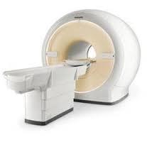 PHILIPS Ingenia 1.5T CX
CT(Computed Tomography)
Our hospital has installed a Siemens 128-slice CT scanner. The device is equipped with an “Adaptive Dose Shield” X-ray exposure reduction mechanism, which blocks unnecessary X-ray exposure for image reconstruction minimizing the exposure of the patient. In addition to the 64-channel multi-slice CT system for the whole body, we have also introduced a high-performance workstation, SYNAPSE VINCENT manufactured by Fujifilm.
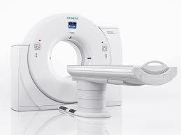 SIEMENS SOMATON Definition AS+
 SYNAPSE VINCENT (3D image analysis)
Mammography
Mammography (breast X-ray examination) is a test to check for cancer when a lump or skin twitch is found by palpation of the breast. An X-ray is taken by holding the breast in a machine. At our hospital, we have introduced the latest mammography equipment with a tomosynthesis function to image subtle density differences in soft tissues such as the breast. High-quality mammography images can be taken with low radiation exposure, which is useful for the detection and treatment of early-stage mammary gland diseases such as microcalcifications.
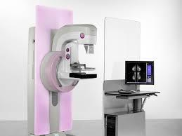 SIEMENS MAMMOMAT Inspiration
Surgical radiography
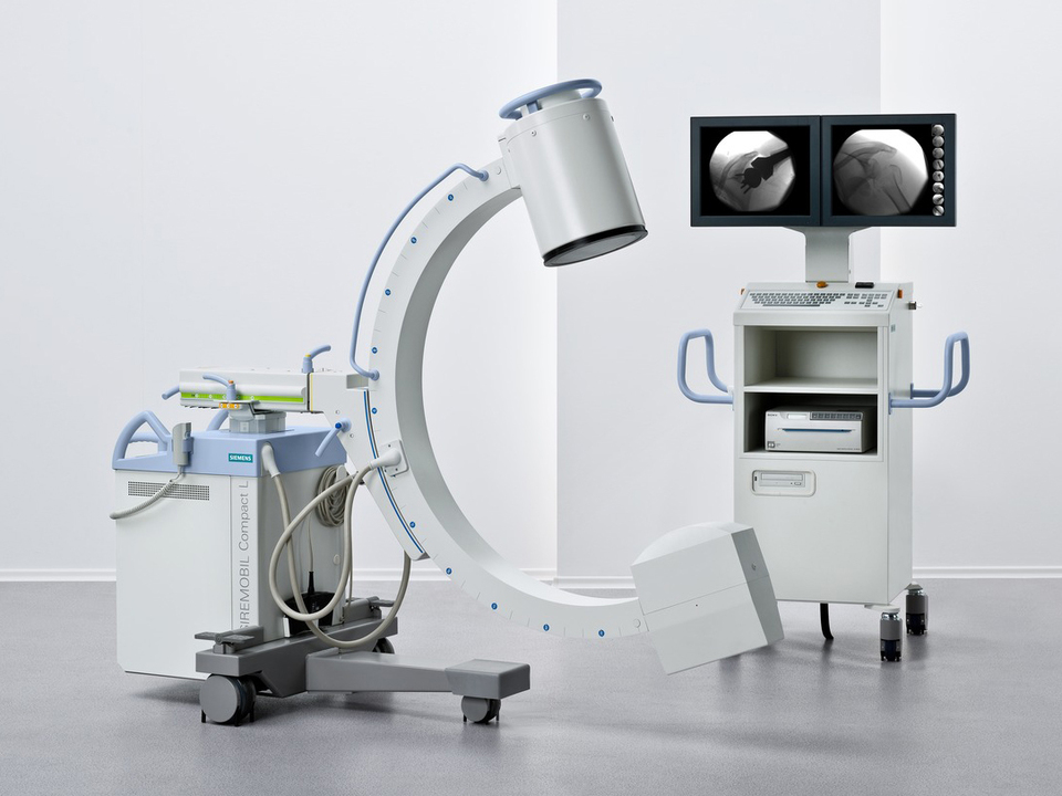
SIEMENS
SIREMOBIL Compact L
Surgical X-ray equipment is a mobile X-ray fluoroscopy and imaging equipment. It is mainly used for orthopedic surgery, and the fixation position during surgery can be confirmed with a fluoroscopic image. It is characterized by a C-arm shape that can be used on an operating table.
X-ray television fluoroscopy
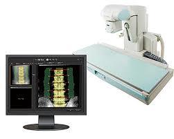
Shimadzu
SONIALVISION G4
Television equipment enables real-time fluoroscopic observation, and typical examinations are to see the flow of barium in the stomach and intestines. In addition, it is a multi-purpose device, such as contrast imaging examination of urology, reduction of fractures and dislocations, and confirmation of the position of the endoscope camera in the body.
Bone strength (measurement of bone mineral content) can be examined to predict the risk of fracture due to osteoporosis.
Angiography
With dramatic progress in IVR using angiography equipment (treatment by applying angiography technology without surgery), angiography is used to treat cerebrovascular diseases such as cerebral aneurysm, cerebral arteriovenous malformation, and cerebral infarction, as well as hyper-selective intra-arterial injection of anti-tumor drugs for abdominal tumors and embolization. Angiography is also used for treatment using balloons and stents (metals for widening) to widen narrowed parts of the blood vessels of the heart. The equipment consists of one biplane type angiography system and one multi-axis angiography system suitable for a hybrid operating room environment.
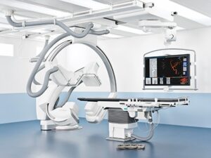 SIEMENS Artis Q BA Twin
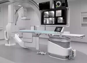 Philips Azurion 7 B12
General radiography
General radiography is the most commonly performed examination in the radiology department, and upon request, images are taken of various parts of the body, such as the chest, abdomen, hands, and feet. Since the examination time is relatively short and the amount of information is large, it is one of the indispensable examinations for the initial diagnosis of various diseases. Our hospital has introduced an FPD (Flat Panel Detector) that can perform inspections with less X-ray dose and obtain high-definition photographs compared to the conventional CR system.
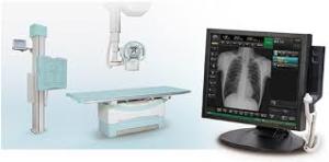 Shimadzu RADSpeed Pro
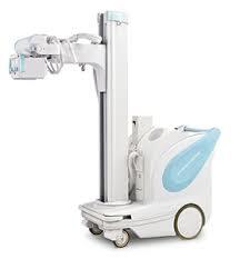 Shimadzu MobileArt Evulution
Ultrasound imaging examination
Ultrasound imaging is a simple and safe test that uses high-frequency sound (ultrasonic waves) that cannot be heard from the outside of the body and uses its reflection to image and observe internal organs(liver, gallbladder, pancreas, kidneys, spleens, thyroid, mammary glands, etc.).
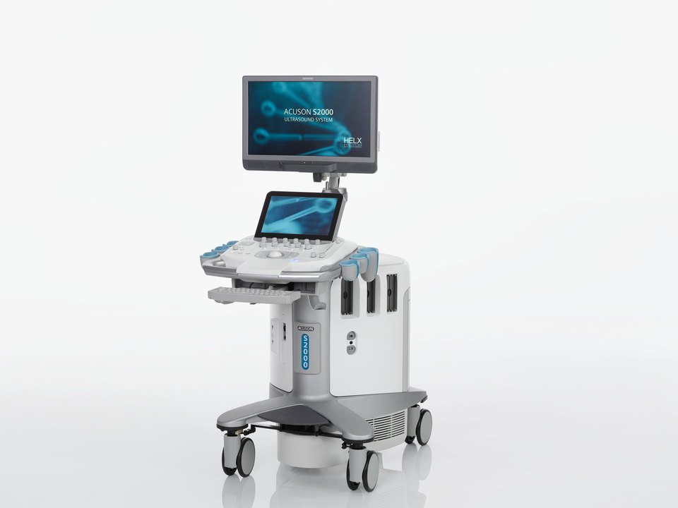 SIEMENS ACUSON S2000 HELX EVOLUTION
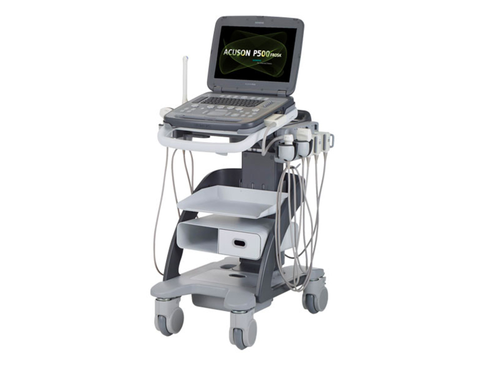 SIEMENS ACUSON P500